The Bad
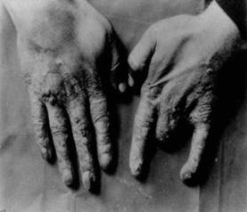
Throughout the years since the first X-rays were made, there have been many incidences where the X-rays would burn the patient’s skin and where the X-rays would not provide a clear image. In July of 1897, Miss Josie MacDonald found a burn on her jaw which later turned out to be necrosis which is a disease decay the bones. This was due to Miss MacDonald being exposed twenty minutes to X-rays while she was getting “an X-ray photography of her upper left jaw” (Burned by the X-Rays). Scientists began to research new methods of improving the X-rays where it would not harm the patient and give a better X-ray image. The New York Times in April 12, 1908 announced that Dr. Albert Geyser, a professor at Cornell Medical College, invented the Cornell tube that “eliminate[ed] danger in the use of X rays” (New Tube Robs X-Ray of Danger). The tube was tried five thousand times and did not burn any patients.
The Good
Through the development of X-rays, many lives can be saved. In November of 1896, Dr. W.Tod Helmuth Jr. was able to find the bullet inside Henry Surgerhelm’s body by using an X-ray. The X-ray showed that the bullet was located “beneath the ribs about an inch and a half above the wound” (Practical X-Ray Work). Without the X-ray, the surgery would have been difficult. In January of 1897, the X-ray saved Thomas Saltmann’s life. For the past years, Saltmann had an affection of the arm which caused him great pain. Before the invention of the X-ray, the doctors were unable to diagnose the patient. However after the invention of the X-ray, the cause of the pain was found. His “deposits of lime salts in the blood [had] hardened the artery” (Photograph of an Artery). Without this invention of the X-ray, these people would have died or would have experienced great suffering. However, thanks to this amazing X-ray, many were saved. Several cases have shown that the Cornell tube also cures patients. In one instance, a “patient…had been cured of epithelioma of the nose after twenty applications of rays from the Cornell tube, [which] ‘is made of heavy lead glass so that no rays are emitted, except through a flint glass window’” (New Tube Robs X-Ray of Danger).
X-rays not only save lives, but it also provide the “means of exploration” (The X-Rays and Exploration). “The growth of bones, from birth till matured age, could be studied with their aid, and the various causes which retard growth (rachitism, tuberculosis) or produce midgets could be ascertained” (The X-Rays and Exploration). X-rays prevent people from being buried alive. It is found that the viscara of a living person is invisible and that “the abdominal organs are in constant movement, and so leave no trace on the photographic plate. In the radiograph of a dead person, on the contrary, the stomach and intestines are clearly marked – this being the case even when the radiograph is taken only a few minutes after death” (Sure Test of Death). X-rays, as seen above, have many implications. Further improvements are being made upon the X-ray and the astonishing development is yet to come.
After:
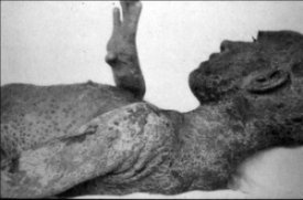
One of Dr. Rollins' patients with dermatitus herpetiformus, before and after X-Ray treatmentCopyright Radiology Centennial Inc. X-Ray technicians fell victim to the horrible side-effects of radiation.
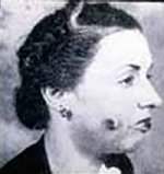
This woman, like thousands of others, suffered
from burns, scarring, and cancer after
undergoing X-Ray "beauty treatments".
Copyright Radiology Centennial Inc
The Ugly
Scientists who have been working with X-rays such as Dr. William Z. Bennett, University of Wooster, has tested X-rays on himself and burned his breast. The tissues and bones of the breast had decayed that the doctors will have to remove part of his breast bone. In June 7, 1915, The New York Times published an article stating that Charles H. Stanley discovered a “new electrical ray”, also known as the Stanley ray, that “does the work of the X-ray more efficiently and without harmful effects” (New Harmless Ray To Do X-Ray’s Work). Stanley’s ray does not harm any patients. It eliminates the fear of patients having to suffer from severe burns. These rays also provide surgeons with a more efficient way of operating people. Instead of having to take “preliminary photographs”, the surgeon is able “to lean over his operating table and see at once…the bullets or bits of shrapnel in the patient’s body” (New Harmless Ray To Do X-Ray’s Work).
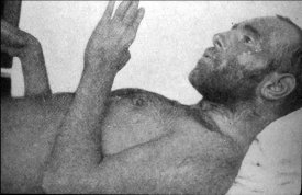
Mihran Kassabian documented and photographed his degeneration,
hoping to help later technicians and patients avoid his fate.
Copyright Radiology Centennial Inc.